
Magnetic resonance imaging (MRI) has changed the way medical professionals diagnose injuries and conditions. This non-invasive imaging technology provides detailed views of soft tissues, bones, and organs, and it enables accurate diagnosis without surgical procedures. Understanding how MRI works and what to expect can help prepare patients for this type of scan.
What Is an MRI?
An MRI is a medical imaging technique that uses magnetic fields and radio waves to create detailed images of the body’s internal structures. Unlike X-rays or CT scans, MRI does not use ionizing radiation, making it a safe option for repeated imaging. The technology produces high-resolution images of soft tissues; this is particularly useful for examining muscles, ligaments, tendons, and the brain.
How Does It Work?
The MRI process relies on the principle of nuclear magnetic resonance. Protons respond to magnetic fields and radio frequencies. When placed in the MRI scanner’s magnetic field, these atoms align in a specific direction, and short bursts of radio waves temporarily knock them out of alignment.
As the protons realign, they release energy that the scanner’s detectors capture. Different tissues release energy at different rates, and this allows the MRI to distinguish among tissue types. A computer processes these signals and constructs detailed cross-sectional images of the body. Radiologists examine these images from multiple angles to identify abnormalities or injuries.
Why Is It Conducted?
These scans are conducted to diagnose a wide range of medical conditions and injuries. Physicians may order MRI scans when they need detailed images of soft tissues that other imaging methods cannot provide. Common reasons include evaluating joint injuries, diagnosing torn ligaments or tendons, and detecting abnormalities in the brain or spinal cord. The technology is particularly valuable in sports medicine because accurate diagnosis of musculoskeletal injuries is needed for proper treatment.
What Does the Process Involve?
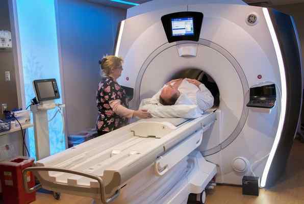
Before the scan, patients may complete a screening form to identify any contraindications. Metal objects must be removed, as the magnetic field can interfere with pacemakers, cochlear implants, and certain metal implants. Patients wear a hospital gown and lie on the examination table, which slides into the scanner. The scanning process can take anywhere from 45 minutes to an hour, depending on the area being examined.
What Are the Benefits?
MRI technology offers several advantages over other imaging methods. The lack of ionizing radiation makes it safe for repeated use, and the technology is suitable for patients who require multiple scans over time. The high level of detail in MRI images allows physicians to detect subtle abnormalities that other imaging techniques may miss.
Another benefit is MRI’s versatility in examining different body systems. A single imaging technology can assess joints, organs, blood vessels, and the nervous system. This reduces the need for multiple scan types, and it streamlines the diagnostic process.
The non-invasive nature of MRI means patients avoid the risks associated with exploratory surgery. Accurate diagnosis through MRI can lead to more targeted treatment plans and better patient outcomes. Early detection of conditions like tumors or vascular abnormalities can improve prognosis and treatment success rates, so consulting a specialist is beneficial.
Learn More About Injury Diagnosis
MRI technology is key to accurate injury diagnosis. The combination of safety, accuracy, and versatility makes it a valuable tool for medical professionals across specialties, and understanding the process helps patients prepare for the procedure. If you have been referred for an MRI scan, speak with your healthcare provider about any questions you may have.