
Neuroradiology plays a significant role in modern healthcare, providing a detailed view of the brain and its intricate network of blood vessels. This field utilizes advanced imaging techniques to identify potential problems before they escalate into serious health events, such as a stroke. By creating precise images of the brain, spinal cord, head, and neck, neuroradiologists can spot abnormalities that might otherwise go unnoticed. This early detection provides a window of opportunity for you and your healthcare team to take preventive action.
How Are At-Risk Areas Identified?
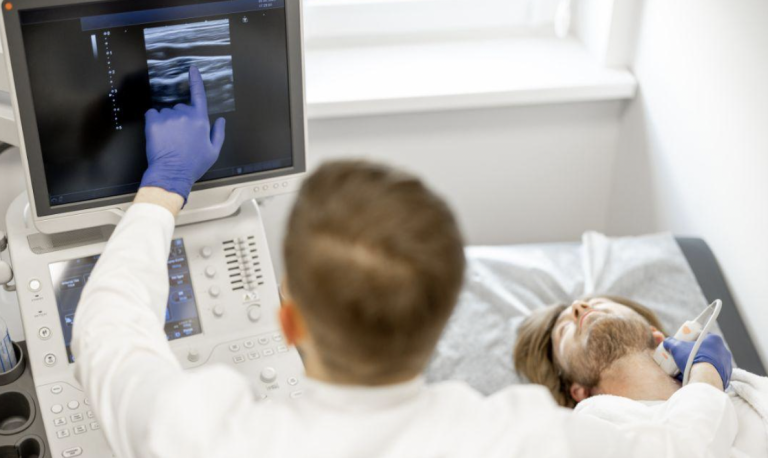
Neuroradiologists use advanced imaging techniques to assess stroke risk by examining brain tissue and blood vessels. These non-invasive methods are pivotal for identifying potential issues before they lead to serious health concerns.
Here are some common imaging tools and what they reveal:
- Magnetic Resonance Imaging (MRI): Produces detailed images of brain tissue to detect abnormalities or damage.
- Computed Tomography (CT) scans: Helps identify blockages or bleeding in the brain.
- Magnetic Resonance Angiogram (MRA): Focuses on arteries and veins, showing areas with plaque buildup or weakened vessel walls.
- CT Angiogram (CTA): Highlights narrowing of arteries (stenosis), aneurysms, or vascular malformations.
These imaging methods give doctors a clear map of your cerebrovascular system, helping them pinpoint at-risk areas. Early detection enables the development of a proactive plan to reduce the risk of stroke and maintain vascular health.
How Do Findings Guide Treatment?
Once imaging identifies a potential risk, the findings help guide the next steps in your care plan. The detailed images from neuroradiology allow your physician to understand the exact location, size, and nature of the issue. This information helps determine the most appropriate course of action, which may range from lifestyle adjustments to medical procedures. Your doctor can develop a personalized approach based on this specific diagnostic information.
The clarity provided by these scans supports informed decision-making between you and your healthcare provider. For instance, the images might show that an artery is only mildly narrowed, suggesting that medication and lifestyle changes could be effective. If the imaging reveals a more significant blockage or a large aneurysm, your medical team will have the precise details needed to plan a more direct intervention. This tailored approach is based directly on what the neuroradiology scans uncover.
When Should Screening Be Discussed?
Discussing screening with your doctor is necessary if you have risk factors for stroke. These factors can help your doctor determine if neuroradiology screening is necessary.
- Family History: A family history of stroke or aneurysm.
- Personal Health Conditions: High blood pressure, diabetes, high cholesterol, or a history of smoking.
- Specific Symptoms: Unexplained severe headaches, dizziness, or temporary neurological symptoms like numbness or weakness on one side of the body.
Bringing these experiences to your doctor’s attention allows them to assess your situation. They can then decide if imaging is needed to investigate the underlying cause and evaluate your stroke risk.
Visit a Neuroradiology Center
Understanding your personal risk for stroke is a key part of managing your long-term health. The detailed insights from neuroradiology can provide you and your doctor with valuable information. If you have risk factors or symptoms that concern you, discussing them with your healthcare provider is a positive first step. They can help you determine if a visit to a neuroradiology center is the right choice for your situation. Taking this proactive step can give you the clarity needed to protect your brain health for the future.