Medical imaging helps transform the way orthopedic spine surgeons approach back problems. Advanced technology allows these specialists to see beyond surface symptoms and understand the root causes of patient discomfort. This detailed visualization facilitates accurate diagnosis and enables the creation of effective treatment plans. Here are a few ways orthopedic spine surgeons use imaging to diagnose back conditions:
Spine Imaging Basics
Orthopedic spine surgeons use imaging to study the complex structures of the back, including vertebrae, discs, ligaments, and surrounding tissues. While these components work together to support movement and protect the spinal cord, the source of discomfort is not always clear during a physical exam. Imaging enables surgeons to examine beyond the surface and pinpoint where problems originate.
Modern technology enables detailed views of the spine from multiple angles, allowing orthopedic surgeons to distinguish between natural, age-related changes and conditions that can require closer evaluation. This added clarity helps provide the context necessary to understand what contributes to back concerns and guides the next steps in patient care.
Diagnostic Tools Explained
Different imaging tools examine the spine, and each highlights specific structures. X-rays can provide a clear view of bone alignment, fractures, and spinal curvature. This information helps determine whether vertebral changes contribute to discomfort.
Magnetic Resonance Imaging (MRI) provides detailed views of soft tissues, including discs, ligaments, and nerve roots. MRI uses magnetic fields and radio waves to produce cross-sectional images. These scans reveal disc herniation, spinal narrowing, or nerve pressure that X-rays might not show.
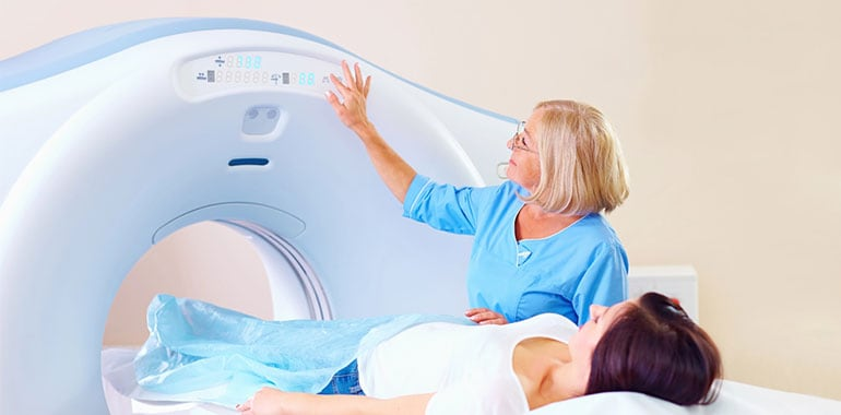
Computed Tomography (CT) combines X-rays with computer processing to create precise cross-sectional views for assessing complex bone issues. Specialized techniques, such as myelography, use contrast material to outline the spinal cord and nerve roots. These methods provide enhanced detail when standard imaging does not fully explain the source of symptoms.
Surgeons Using Imaging
Orthopedic surgeons study imaging results so they can link structural spine changes with reported symptoms. Interpreting these images requires training, as normal variations sometimes resemble abnormalities. Specific patterns are recognizable, such as a disc pressing on a nerve root, which has a distinct MRI appearance.
Image interpretation is combined with physical examination and patient history to create a diagnostic picture. This process distinguishes between changes that cause symptoms and those that do not. Detailed imaging also supports surgical planning when non-surgical options have not resolved the issue.
Back Condition Insights
Imaging helps clarify a range of spinal conditions by showing distinct patterns and structural changes. These images make it easier to understand which areas are affected and the severity of the issue. Common examples include:
- Herniated discs: Bulging or ruptured disc material pressing on nearby nerves, with clear visibility of affected levels.
- Spinal stenosis: Narrowing of the spinal canal caused by bone spurs, thickened ligaments, or enlarged joints.
- Compression fractures: Breaks from trauma or osteoporosis, with images distinguishing recent injuries from older, healed ones.
- Degenerative changes: Gradual developments such as disc space narrowing, bone spurs, and joint wear are linked to long-term discomfort.
Find an Orthopedic Spine Surgeon Today
Imaging results guide orthopedic spine surgeons in creating tailored treatment plans that can address both function and quality of life. Some findings support conservative care, such as physical therapy or medication, while more severe conditions may point toward surgical correction. These images act as a roadmap, clarifying which structures need attention and how to proceed safely. Follow-up imaging also helps monitor healing and refine care when needed. If you’re navigating back concerns, consider asking your provider how imaging fits into your treatment plan.